

Web Desk: Scientists are making steady progress toward a long-sought goal in hearing research after discovering that the mammalian inner ear may retain a hidden ability to regenerate critical sensory cells, raising hopes for future treatments for permanent hearing loss.
For years, researchers believed that the loss of sensory hair cells inside the inner ear was irreversible. These specialized cells, which convert sound vibrations into electrical signals for the brain, do not naturally regenerate in humans once they are damaged by aging, prolonged noise exposure, disease, certain medications or genetic conditions.
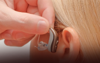
Although hearing aids amplify sound and cochlear implants help bypass damaged structures, neither treatment restores the lost cells responsible for hearing.
By contrast, several non-mammalian species, including birds and fish, can naturally replace damaged sensory hair cells, allowing them to recover hearing after injury.
However, research over the past decade has challenged the long-standing belief that mammals completely lack regenerative capacity.
Scientists found that newborn mice possess a brief period after birth during which they can regenerate lost sensory hair cells. That ability fades rapidly within days, suggesting mammals may not have entirely lost the biological machinery needed for regeneration but instead lose access to it as they mature.
The discovery marked a significant turning point in hearing research.
“Prior to that, the dogma was regeneration only happens in non-mammals,” said Brandon Cox, a developmental biologist at Southern Illinois University School of Medicine.
The findings prompted researchers to shift their focus from searching for an entirely new mechanism to understanding how dormant regenerative programs might be reactivated.
Instead of identifying a single genetic switch capable of restoring hearing, scientists now believe regeneration depends on activating a combination of developmental signals.
Their research has highlighted an unexpected source of replacement cells: supporting cells that surround sensory hair cells within the cochlea.
Under the right molecular conditions, these supporting cells can begin transforming into hair cell-like structures. Researchers are now working to help the cells complete that conversion so they function like fully mature sensory hair cells.
Hearing begins inside the cochlea, a spiral-shaped organ located deep within the inner ear.
The cochlea contains about 15,000 sensory hair cells, each topped with microscopic structures known as stereocilia. As sound waves enter the ear, these tiny projections bend, triggering electrical signals that travel through the auditory nerve to the brain, where they are interpreted as sound.
The process is highly sensitive. Years of exposure to loud noise, aging, chemotherapy drugs, certain antibiotics and inherited genetic mutations can permanently damage these cells.
Unlike tissues such as skin, blood or the liver, the mammalian cochlea has little natural ability to replace lost sensory cells. Once hair cells die, neighboring supporting cells typically remain inactive instead of generating replacements.
Researchers believe the answer may lie partly in epigenetics, the biological system that controls which genes a cell can access and activate.
According to Cox, young cells have more open DNA, allowing developmental genes to function more easily. As cells mature, the surrounding chromatin becomes increasingly compact, making those same genetic instructions far less accessible.
As a result, the genes required to create new hair cells remain present in adult tissue but are effectively locked away behind layers of molecular regulation.
Scientists are now investigating ways to reopen those developmental pathways and encourage mature cells to behave more like their younger counterparts.
The search has identified three key transcription factors Atoh1, Gfi1 and Pou4f3 that play central roles during embryonic development by directing immature cells to become sensory hair cells.
Transcription factors regulate networks of genes, determining which genetic instructions are switched on or off as cells develop specialized functions.
Earlier research focused largely on Atoh1 because it acts as one of the earliest signals involved in hair cell formation. Scientists hoped reactivating the protein in damaged ears would stimulate the growth of new sensory cells.
Although experiments produced cells expressing some hair cell characteristics, many remained immature. They often failed to develop fully formed stereocilia, lacked the specialized features of mature inner and outer hair cells and did not establish the neural connections necessary for hearing.
Nevertheless, the work provided an important proof of concept and opened new avenues for gene therapy research.
“It opened the road for others to consider gene therapy,” said Alan Cheng, a surgeon and professor of otolaryngology at Stanford University.
Researchers now believe successful hearing restoration will require coordinating multiple developmental signals rather than relying on a single genetic trigger, bringing the field closer to understanding how damaged hearing may one day be repaired.
Read more: ChatGPT announces major cuts in API prices